
Cathepsin G (CATG) is a core serine protease of neutrophils. It is extensively involved in pathophysiological processes such as inflammatory immunity, vascular remodeling and tumor microenvironment regulation, making it a vital target for functional detection in scientific research. It should be noted that protein expression levels cannot reflect enzymatic functional activity. Only the measurement of CATG enzymatic activity can truthfully mirror the functional status of samples, validate experimental phenotypes and assess the intervention effects of drugs, which is also the key to eliminating experimental data bias and irreproducible results.
Traditional colorimetric assays have inherent drawbacks including low sensitivity, substantial detection errors for trace samples and incompatibility with high-throughput tests. By contrast, the fluorometric assay for CATG enzyme activity has emerged as the mainstream detection protocol owing to its merits of high sensitivity, low background signal, real-time quantification and straightforward operation.
Nevertheless, numerous researchers frequently encounter issues including intense background fluorescence interference, fluctuating readings and poor data repeatability during practical operations, resulting from improper reaction system ratios, uncontrolled incubation parameters and non-standard control groups. Such troubles waste considerable time and samples yet fail to yield valid experimental data.
To address the above common experimental challenges, this issue compiles a complete, ready-to-implement practical protocol for fluorometric CATG enzyme activity detection. Novice researchers may apply the protocol directly, while experienced investigators can optimize the reaction system based on it to reliably complete CATG enzyme activity measurement.


Cathepsin G (CATG) specifically recognizes and hydrolyzes the fluorogenic substrate MeOSuc-Ala-Ala-Pro-Met-AMC by precisely cleaving the amide bond between methionine (Met) and the AMC fluorophore.
Fluorescence of intact MeOSuc-Ala-Ala-Pro-Met-AMC is intrinsically quenched. After enzymatic hydrolysis, free 7-amino-4-methylcoumarin (AMC) is released into the reaction solution and generates characteristic detectable fluorescence. As CATG continuously hydrolyzes substrate molecules over the incubation period, the concentration of free AMC rises linearly, accompanied by a linear increase in fluorescence intensity over time.
The catalytic activity of CATG is directly proportional to the rate of fluorescence signal elevation. Under identical detection parameters, a faster increase in fluorescence intensity corresponds to higher CATG concentration / enzymatic activity within the sample. CATG activity can be quantified by measuring the growth rate of fluorescence signals.
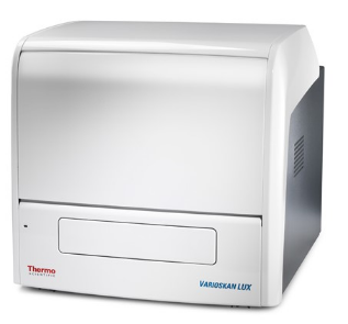
Main Instruments and Reagents
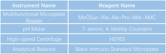

1. Preparation of working buffer (CATG Assay Buffer): Prepare 100 mM HEPES solution and adjust pH to 7.5.
2. Tissue pretreatment: Weigh approximately 0.1 g tissue sample, add 1 mL lysis buffer for tissue lysis. Centrifuge the mixture at 10,000 rpm for 10 min at 4 °C. Collect the supernatant as crude enzyme extract and keep it on ice for subsequent detection.
3. Preparation of CATG fluorogenic substrate solution: Thaw 80 mM CATG fluorogenic substrate stock solution at room temperature, then dilute it with CATG Assay Buffer to prepare 200 μM CATG fluorogenic substrate working solution.
4. Preparation of CATG enzyme standard: Verified fresh mouse liver homogenate can be used as CATG enzyme standard. Weigh and homogenize fresh mouse liver tissue, followed by centrifugation to collect the supernatant of liver homogenate. Determine the appropriate concentration via preliminary testing, aliquot and store at -80 °C; repeated freeze-thaw cycles shall not exceed once.
5. Quantification of molar amount corresponding to unit AMC fluorescence: Thaw AMC stock solution at room temperature, dilute it with CATG Assay Buffer to prepare serial AMC standard solutions according to the corresponding table. Vortex thoroughly after each dilution step before proceeding to the next gradient dilution.

6.Pipette AMC standards and AMC-blank control into the Black Immuno Standard Microplate sequentially at a volume of 100 µL per well.
7.Read the plate using the fluorescence detection module of the multifunctional microplate reader with excitation wavelength set at 380 nm and emission wavelength at 460 nm. Perform linear regression with AMC fluorescence intensity as the y-axis and AMC concentration as the x-axis, then calculate the intercept (a), slope (b) and correlation coefficient (\(r^2\) > 0.99) of the regression curve.
Calculation formula for molar quantity of AMC per relative fluorescence unit (RFU):
Molar amount of AMC per RFU (pM/RFU) = \(x × 100 µL ÷ 1000000 ÷ Y\)
8.Sample detection:Reader parameter setup: Kinetic measurement for 30 min, plate reading interval of 30 s, incubation temperature at 37 °C, excitation wavelength 380 nm, emission wavelength 460 nm.
Add samples, CATG enzyme standard and blank control (CATG Assay Buffer) sequentially into the Black Immuno Standard Microplate at 50 µL per well. Then supplement each well with 50 µL of 200 μM CATG fluorogenic substrate working solution. Shake the plate gently for uniform mixing and immediately place it into the microplate reader for fluorescence recording.
9.Calculation of enzymatic activity:CATG activity of test sample (pM/min) = Vmax of test sample × molar amount of AMC per RFU (pM/RFU)

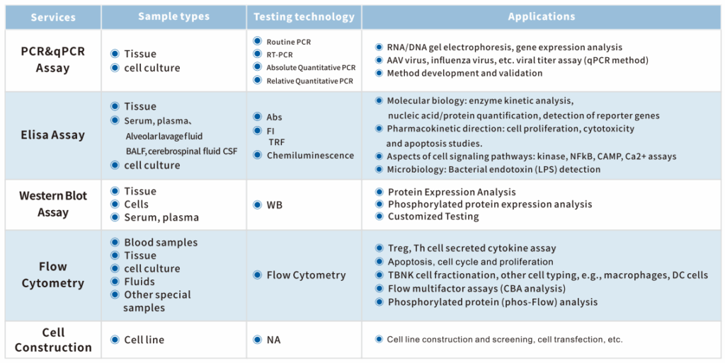
Equipped with cutting-edge technical equipment and abundant project experience, the KCI・KMQ Molecular Biology Platform can undertake biochemical detection assays with a daily throughput of over 300 samples. Meanwhile, it provides customers with a full spectrum of molecular biology testing services, including flow cytometry analysis, qPCR, ELISA, Western Blot (WB), IP and Co-IP. At present, our company has established extensive long-term cooperative relationships with numerous well-known pharmaceutical enterprises and scientific research institutions at home and abroad, laying a solid technical foundation for the research and development of innovative drugs.